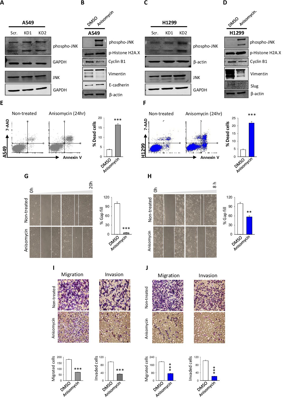
Fig. 5. JNK activation is contributing to TRPM2 KD-mediated NSCLC death and invasion-blocking. (A & C) Western blot analysis of JNK and pJNK protein level in control and TRPM2-KD cells (B & D) Protein expression level of pJNK, p-Histone H2A.X, Cyclin B1 and vimentin in NSCLC control cells, with and without treatment with 1 μg/mL Anisomycin for 24 hrs (E & F) Annexin V/7-AAD staining of apoptotic and necrotic cells in both Anisomycin (1 μg/mL Anisomycin for 24 hrs) treated and non-treated cells; percentages of dead cells are presented as bar graphs (G & H) Gap closure analysis of cell motility in the presence and absence of 1 μg/mL Anisomycin (I & J) The quantitative analysis of migration and invasion ability of Anisomycin treated and non-treated cells after 16 hrs incubation (t test vs scr. ***, p<0.001; **, p<0.01; * p<0.05).